Immunofluorescence Protocol Frozen Section

Graphic Protocol For The Preparation And Fluorescent Ihc Staining Of Frozen Tissue Sections R D Systems
Immunohistochemistry Leinco Technologies

Frozen Section Staining For Immunofluorescence If Microscopy Institute For Molecular Bioscience University Of Queensland
Ihc Protocols Antibody Company Buy Antibodies Online

Cryogenic Tissue Processing And Section Immunofluorescence Of Cerebral Organoids
Considerations For Immunofluorescence Staining Biotium

Protocol for immunofluorescent staining of mouse frozen sections tissue.
Immunofluorescence protocol frozen section.

Immunofluorescence Test An Overview Sciencedirect Topics

2017 Fluomute Ready To Use Reagent To Reduce Autofluorescence In Cells And Tissue Just Incubate Fixed Cells Of Tissue Cells And Tissues Stem Cells Treatment
Watch Video Immunofluorescence Tissue Staining Biocompare Streaming Video

A Manual Multiplex Immunofluorescence Method For Investigating Neurodegenerative Diseases Biorxiv
Immunofluorescence Troubleshooting Protocol Tips Stressmarq
Free Floating Or Slide Mounted Immunohistochemistry

Overview Of Immunohistochemistry Thermo Fisher Scientific Ca
Ly6g Antibody Rb6 8c5 Ab25377 Abcam
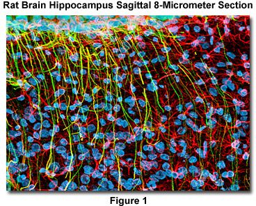
Confocal Microscopy Staining Brain Cryosections Olympus Life Science
Mouse Phenotyping Ucsd University Of California San Diego

580 Questions With Answers In Immunofluorescence Staining Scientific Method

Recombinant Anti Tbr1 Antibody Epr8138 2 Ab183032 Abcam

Pin On Moj Toxicology

Frozen Tissue Slide Preparation And Processing Youtube

Recombinant Anti Tmem119 Antibody 28 3 Microglial Marker Ko Tested Ab209064

Astrocyte Marker Aldh1l1 Eaat1 Eaat2 Gfap Antibody Sampler Panel Ab226481
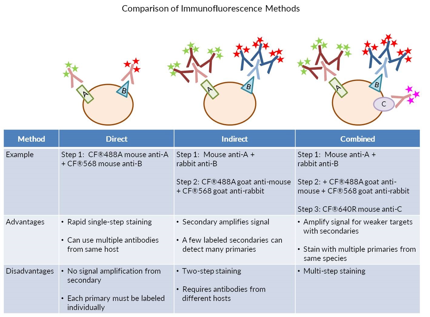
Our Comprehensive Guide To Performing Double Immunofluorescence

Mounting Medium With Dapi Aqueous Fluoroshield Ab104139 Abcam
Https Encrypted Tbn0 Gstatic Com Images Q Tbn 3aand9gcsovvalcw Lhxvcxg6jl7eq7c5qqa7spkadlw0ad27m1gwojhyi Usqp Cau
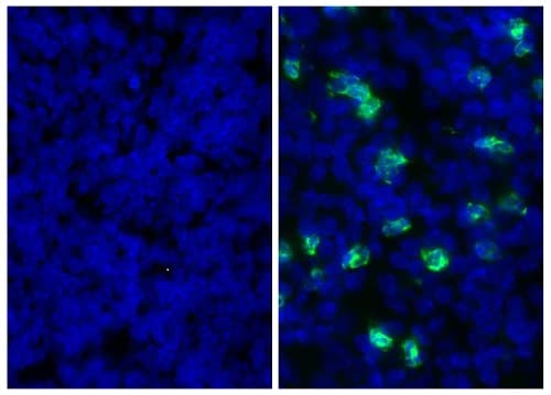
Anti Brdu Antibody Bu1 75 Icr1 Proliferation Marker Ab6326 Abcam

Anti Map2 Antibody Ab5392 Abcam

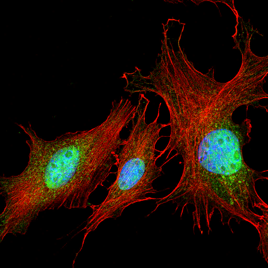
Collagen I Antibody Nb600 408 Novus Biologicals

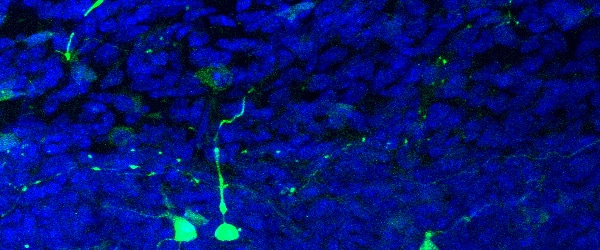
Immunofluorescence Neun Staining Of Frozen Mouse Brain Sections Mainly Staining The Cytoplasm And Not The Nucleus Of Neurons

Ihc Protocol Paraffin Frozen And Free Floating Sections Abcam
Source : pinterest.com
